Bones In Leg Diagram - Leg Bone Diagram Anatomy Bones Physiology Medical Anatomy
Bones In Leg Diagram - Leg Bone Diagram Anatomy Bones Physiology Medical Anatomy. The proximal portion of the tibia is tibial plateau which acts as a cusp for the knee, the distal portion tapers into the medial malleoli and the concave surface which articulates with the talus at the ankle joint. The talus is the bone at the top of the foot. The knee joint is the largest joint in the body and is primarily a hinge joint, although some sliding and rotation occur. It connects with the tibia and fibula bones of the lower leg. Learn vocabulary, terms, and more with flashcards, games, and other study tools.
ads/bitcoin1.txt
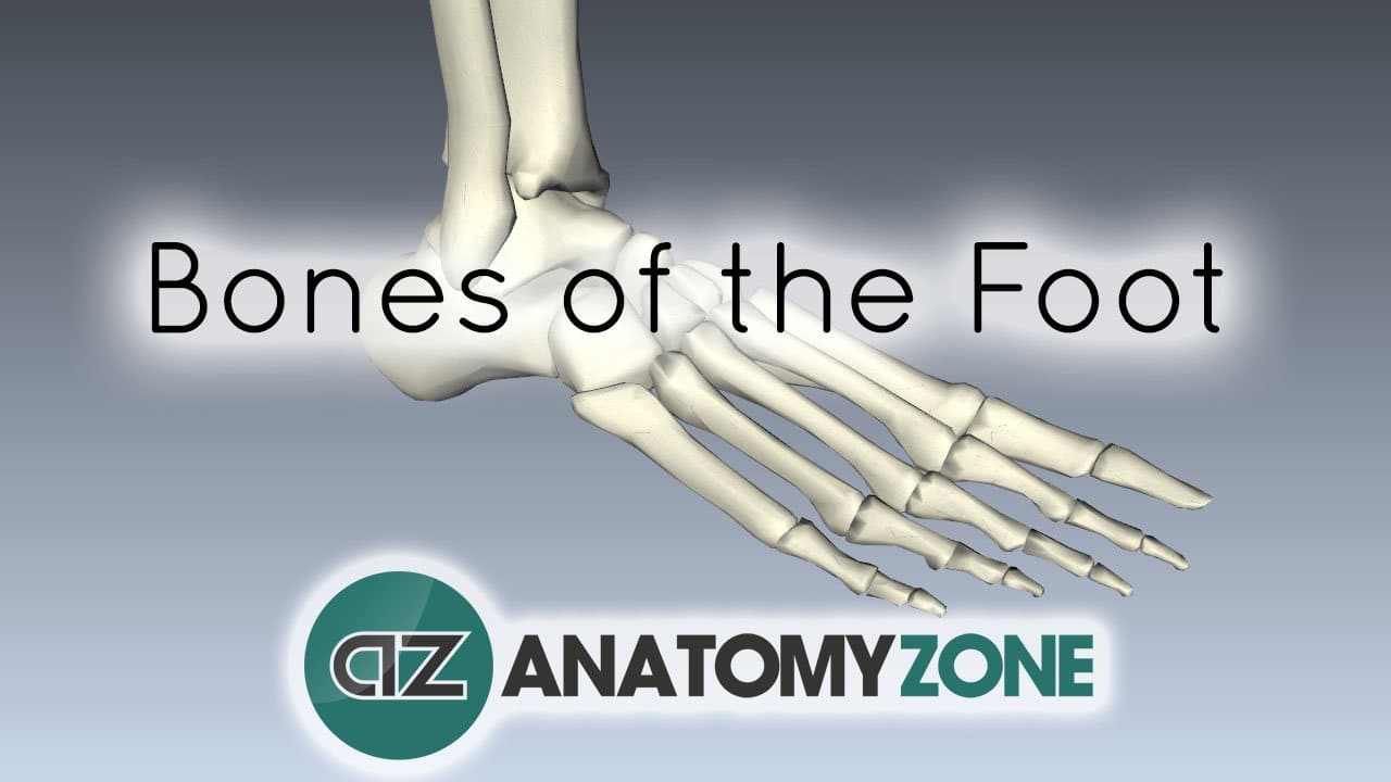
The upper arm and the forearm. The femur is the single bone of the thigh. The tibia, commonly known as the 'shin bone', is the largest and most medial of the two.you can palpate its anterior border when you run your finger down the anterior aspect of your leg. The bones of the leg are the femur, tibia, fibula and patella.the foot bones shown in this diagram are the talus, navicular, cuneiform, cuboid, metatarsals and calcaneus. These landmarks are the anterior superior iliac spine.
Horse Anatomy Diagrams The Anatomy Of A Horse Horse Anatomy Animal Medicine Horse Care from i.pinimg.com Also called the shin bone, the tibia is the longer of the two bones in the. The smaller lateral bone of the lower leg. Learn the muscles of the leg fast with these quizzes, diagrams and labeling exercises : The upper arm and the forearm. (note, the radius and ulna bones also have this membrane.) this membrane keeps the tibia and fibula together and provides strength and stability for them. The major bones of the leg are the femur (thigh bone), tibia (shin bone), and adjacent fibula, and these are all long bones.the patella (kneecap) is the sesamoid bone in front of the knee.most of the leg skeleton has bony prominences and margins that can be palpated and some serve as anatomical landmarks that define the extent of the leg. Bones of the lower limb anatomy and physiology from opentextbc.ca time to jump right into the biggest and strongest bones in the human body. Bone xray ankle anatomy 12 photos of the bone xray ankle anatomy , bone.
Also called the shin bone, the tibia is the longer of the two bones in the.
ads/bitcoin2.txt
Its lower end helps create the knee joint. Leg bone diagram labeled : There are in all 7 bones, which fall under tarsal bones category. Learn vocabulary, terms and more with flashcards, games and other study tools. Quadriceps and hamstring muscles are the major ones which are associated with the knee joint. Bones of the lower limb anatomy and physiology from opentextbc.ca time to jump right into the biggest and strongest bones in the human body. These landmarks are the anterior superior iliac spine. Now let's look at the tibia bone, which is the larger of the two leg bones, located medially. The proximal portion of the tibia is tibial plateau which acts as a cusp for the knee, the distal portion tapers into the medial malleoli and the concave surface which articulates with the talus at the ankle joint. These are the femur, patella, tibia, fibula, tarsal bones, metatarsal bones, and phalanges (see figure 6.51). Four quadriceps muscles are present in front of the knee which help in straightening the leg from the knee. The lower leg extends from the knee to the ankle. The diagram of bones in the ankle and foot is given below:
These muscles work together to produce movements such as standing, walking, running, and jumping. The hip itself is a ball and socket joint, much like the shoulder.the structures necessary to create this joint are the socket, the joint capsule, muscle, ligaments, and the neck. It also separates muscles on the anterior and posterior parts of the leg. Learn vocabulary, terms and more with flashcards, games and other study tools. Quadriceps and hamstring muscles are the major ones which are associated with the knee joint.
Lower Limb 3d Interactive Anatomy Tutorials from anatomyzone.com In total, the bones in the arm are three in total. Quadriceps and hamstring muscles are the major ones which are associated with the knee joint. Leg femur diagram data wiring diagram today. It also separates muscles on the anterior and posterior parts of the leg. Leg pain can also be caused by blood clots, varicose veins or poor circulation. Bone xray ankle anatomy 12 photos of the bone xray ankle anatomy , bone. Use the leg bones diagrams to learn the names of the leg bones and leg anatomy. Bones of the lower limb anatomy and physiology from opentextbc.ca time to jump right into the biggest and strongest bones in the human body.
Posted on april 18, 2019april 18, 2019.
ads/bitcoin2.txt
The foot bones shown in this diagram are the talus, navicular, cuneiform, cuboid, metatarsals and calcaneus. To explain the term in layman's language, it is the heel bone in the skeletal system. With different grades of sprains depending on severity. Start studying pelvis, leg bones, leg bones. Learn vocabulary, terms, and more with flashcards, games, and other study tools. Leg pain can also be caused by blood clots, varicose veins or poor circulation. The tibia and the fibula, at the top of the ankle joint. The lower leg extends from the knee to the ankle. The bones together make up the hip. At the same time, the bones and joints of the leg and foot must be strong enough to support the body's weight while remaining. Bone diagram forehead (frontal bone) nose bones (nasals) cheek bone (zygoma) upper jaw (maxilla) lower jaw (mandible) breast bone (sternum) upper arm bone (humerus) lower arm bone (ulna) thigh bone (femur) collar bone (clavicle) toe bones (phalanges) ankle bones (tarsals) kneecap (patella) shin bone Another bone that is part of the lower leg and the knee joint is called the fibula.this is a bone located on the lateral, or outer part, of the lower leg and is more commonly known as the calf bone. Quadriceps and hamstring muscles are the major ones which are associated with the knee joint.
The talus is the bone at the top of the foot. Leg pain can also be caused by blood clots, varicose veins or poor circulation. Now let's look at the tibia bone, which is the larger of the two leg bones, located medially. Four quadriceps muscles are present in front of the knee which help in straightening the leg from the knee. Many muscles that move the trunk and legs, such as our abdominal muscles, attach to the hip bones.
Foot And Leg Anatomy Essential Info For Yoga Teachers from www.yogajournal.com The tibia and fibula are two long bones that run parallel to each other, forming the scaffold of the leg and providing attachment points for many muscles. Leg bone diagram labeled : The bone in the upper arm is the humerus while ulna and radius make up the forearm. The bones of the hip include the femur, the ilium, the ischium, and the pubis. The patella is the kneecap and articulates with the distal femur. The knee joint is the largest joint in the body and is primarily a hinge joint, although some sliding and rotation occur. The lower limb contains 30 bones. Bone xray ankle anatomy 12 photos of the bone xray ankle anatomy , bone.
Learn vocabulary, terms and more with flashcards, games and other study tools.
ads/bitcoin2.txt
The talus is the bone at the top of the foot. At the same time, the bones and joints of the leg and foot must be strong enough to support the body's weight while remaining. This area is commonly referred to as the calf. The smaller lateral bone of the lower leg. In addition, the broad hip bones provide protection to the delicate internal organs of the pelvis, such as the intestines, urinary bladder, and uterus. The hip itself is a ball and socket joint, much like the shoulder.the structures necessary to create this joint are the socket, the joint capsule, muscle, ligaments, and the neck. Quadriceps and hamstring muscles are the major ones which are associated with the knee joint. The lower leg is comprised of two bones, the tibia and the smaller fibula. The tarsal bones in the foot are located amongst tibia, metatarsal bones, and fibula. Leg bone diagram labeled : Learn vocabulary, terms and more with flashcards, games and other study tools. These are the femur, patella, tibia, fibula, tarsal bones, metatarsal bones, and phalanges (see figure 6.51). The major bones of the leg are the femur (thigh bone), tibia (shin bone), and adjacent fibula, and these are all long bones.the patella (kneecap) is the sesamoid bone in front of the knee.most of the leg skeleton has bony prominences and margins that can be palpated and some serve as anatomical landmarks that define the extent of the leg.
ads/bitcoin3.txt
ads/bitcoin4.txt
ads/bitcoin5.txt
0 Response to "Bones In Leg Diagram - Leg Bone Diagram Anatomy Bones Physiology Medical Anatomy"


0 Response to "Bones In Leg Diagram - Leg Bone Diagram Anatomy Bones Physiology Medical Anatomy"
Post a Comment